August 2003 Newsletter: The tubercle of Zuckerkandl
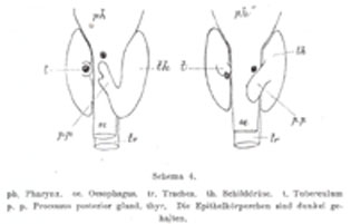
The tubercle of Zuckerkandl is a distinct anatomical entity that was first described by Emil Zuckerkandl in 1902. The description, and the clinical significance of the tubercle, was lost to endocrine surgeons until the last decade when the tubercle was “rediscovered” by a number of authors. Thyroid development involves the midline descent of thyroid tissue from the foramen caecum to the level of the larynx along the thyroglossal tract, at which stage the left and right lobes develop. What is not as well appreciated is that there is, in addition, a lateral thyroid component arising from the 4th branchial cleft and ultimobranchial body which fuses with the median component to form the tubercle of Zuckerkandl. This fusion is not only the source of the thyroid’s C-cells, but is also essential in the process of follicular development. Emil Zuckerkandl studied medicine at the University of Vienna in 1867 and distinguished himself such that eventually, following the death of Langer in 1888, he assumed the Chair in Anatomy at Vienna. He was noted for his sharp observational powers and critical mind making significant contributions to normal and pathological anatomy. He is also one of the very few medical scientists who is remembered as much for his wife, Bertha Zuckerkandl-Szeps, one of the most remarkable personalities of intellectual Jewish society in Vienna during the last decades of the Austro-Hungarian empire. Their house became the meeting point for the avant garde in arts and science, including the sculptor Auguste Rodin and the composer Gustav Mahler. Zuckerkandl’s name is associated with seven eponymous anatomical/surgical entities:
- The Organ of Zuckerkandl – a mass of chromaffin tissue found adjacent to the anterior aspect of the aorta, between the inferior mesenteric artery and the aortic bifurcation
- Zuckerkandl's fascia - the posterior layer of the renal fascia
- Zuckerkandl's gyrus (Zuckerkandl's convolution) - the thin sheet of grey/white substance in front of, and ventral to, the genu of the corpus callosum
- The Concha of Zuckerkandl - a rarely found small nasal concha situated above the supreme nasal concha
- Zuckerkandl's dehiscence - small gaps occasionally seen in the layers of the ethmoid bone
- Zuckerlandl's operation - perineal prostatectomy
- The tubercle of Zuckerkandl – a posterolateral projection of the thyroid gland derived from the 4th branchial cleft
The importance of the tubercle of Zuckerkandl is that, if not looked for and removed during thyroid surgery, it may be a source of persistent unrelieved symptoms or recurrence. An understanding of the anatomy of the tubercle of Zuckerkandl is also central to safe surgical dissection. It usually enlarges lateral to the recurrent laryngeal nerve, with the nerve appearing to pass into a cleft medial to it – a situation that some surgeons used to describe as the nerve passing into the thyroid substance. Early elevation of the tubercle of Zuckerkandl usually allows the recurrent nerve to be easily and safely “encountered” even though not initially visible. However an uncommon but high risk situation is where the recurrent laryngeal nerve runs lateral to an enlarged Tubercle of Zuckerkandl, placing it at increased risk of damage during dissection. Another important point is that the normal superior parathyroid gland, also being derived from the 4th branchial cleft, is commonly found in close association, cephalad to the Tubercle of Zuckerkandl.
